

Most cavities don’t start where you can see them. They start in the tight space between two teeth, exactly where a dentist’s eyes and a mirror can’t reach. That’s the entire reason a dental bitewing x-ray exists. It’s the one imaging tool built specifically to catch decay in the one place a visual exam consistently misses.
A dental bitewing x-ray is a type of intraoral x-ray that captures the crowns of the upper and lower back teeth on one side of the mouth in a single image, showing the space between each tooth along with the bone level just below the gumline. The name comes from the small tab or wing on the x-ray sensor that the patient bites down on to hold it in place. Unlike a full mouth panoramic x-ray, which shows the entire jaw in one wide image, a bitewing zooms in on a specific set of teeth to catch detail that a broader image would blur.
This matters because interproximal cavities, the ones that form between teeth, are one of the most common types of decay and among the hardest to catch early without imaging. A dentist can look directly at a tooth’s visible surface and miss a cavity that’s already spreading beneath the contact point with the tooth next to it. A bitewing x-ray closes that gap. It’s typically taken during a routine checkup, usually as a set of two or four images covering both sides of the mouth, and it’s one of the most common tests performed at a general dental visit in the United States.
Beyond cavities, a bitewing also shows the height of the bone that supports each tooth, which makes it useful for catching early signs of bone loss from gum disease before it becomes visible or symptomatic. It can also reveal problems with old fillings, like a filling that’s started to break down or leak at the edges, well before that shows up as sensitivity or pain.
This guide covers exactly what a dental bitewing x-ray shows, how it differs from other dental x-ray types, how often dentists recommend getting one, what the radiation exposure actually involves, and what to expect during the appointment itself.
What Does a Bitewing X-Ray Show?
Direct answer: A bitewing x-ray shows the crowns of the upper and lower back teeth on one side of the mouth, along with the space between adjacent teeth and the top of the supporting bone. It’s primarily used to detect cavities between teeth, check the condition of existing fillings, and monitor bone loss from gum disease, none of which are reliably visible during a standard visual exam.
Dentists read a bitewing for a few specific things every time:
- Cavities between teeth, which show up as dark shadows or notches at the point where two teeth touch.
- Bone level, since healthy bone should sit close to the base of the tooth, and a gap indicates bone loss.
- The fit and condition of existing fillings and crowns, checking for gaps, overhangs, or decay forming underneath an old restoration.
- Tartar buildup below the gumline, which isn’t visible during a routine visual check but shows clearly on the image.
Dental Bitewing X-Ray vs Other Dental X-Ray Types
Bitewings are just one of several dental x-ray types, and each is built for a different purpose. A panoramic x-ray captures the entire mouth, jaw, and sinuses in one wide image, which makes it useful for evaluating wisdom teeth position, jaw structure, and overall development, but it isn’t detailed enough to reliably catch small cavities between teeth. A periapical x-ray focuses on a single tooth from crown to root tip, which is typically used to investigate a specific problem like an infection or abscess rather than for a routine cavity check. A closer comparison of panoramic radiography versus bitewing imaging breaks down exactly when dentists choose one over the other.
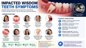
For a wisdom tooth specifically, a panoramic x-ray is generally the better tool for seeing its position and whether it’s impacted, which is covered in more detail in this look at impacted wisdom teeth symptoms. But once a wisdom tooth has erupted enough to sit next to its neighbor, a bitewing becomes useful again, since it’s the image type that can actually catch a cavity forming at that new contact point, a common but easy-to-miss cause of cavity in wisdom teeth pain.
Are Dental X-Rays Safe?
Dental x-rays, including bitewings, use a very low dose of radiation, and modern digital sensors have reduced that exposure significantly compared to older film-based systems. A single bitewing x-ray delivers a small fraction of the radiation involved in many everyday activities, and dental offices in the United States follow protective protocols, including lead aprons and, in many cases, thyroid collars, to minimize exposure further. For a full breakdown of radiation levels across different dental x-ray types and how offices manage exposure, this overview of types of dental x-rays covers the safety measures in more depth. Anyone with specific concerns, including pregnancy, should raise them directly with their dentist before the appointment, since protocols can be adjusted based on individual circumstances.
How Often Should You Get a Bitewing X-Ray?
For most adults with a low cavity risk and no active dental problems, dentists typically recommend bitewing x-rays once every one to two years. Patients with a higher risk of cavities, active gum disease, or recent dental work are often advised to get them more frequently, sometimes every six to twelve months, so a dentist can track changes closely. The right interval depends on individual oral health history, and it’s a decision made together with a dentist rather than a fixed rule that applies to everyone.
What to Expect During a Bitewing X-Ray
The process is quick and typically takes just a few minutes for a full set:
- A small sensor or piece of film, with a tab attached, is placed inside the mouth against the back teeth.
- The patient bites down gently on the tab to hold the sensor in place and keep the teeth aligned in the image.
- The x-ray machine captures the image from just outside the cheek, and the process repeats for each side of the mouth.
- Digital sensors display the image almost instantly, letting the dentist review it during the same appointment.
There’s no discomfort involved beyond the mild sensation of biting on the tab, and no recovery time is needed afterward.
Bitewing X-Rays and Wisdom Tooth Pain
Because wisdom teeth sit at the very back of the jaw, they’re one of the hardest areas to evaluate without imaging. A bitewing can pick up a cavity forming at the contact point between a wisdom tooth and the molar next to it long before any wisdom tooth pain symptoms appear, which matters since decay in that spot can progress silently. This is especially relevant when a cavity develops with no pain at all, since a bitewing is often the only way that kind of decay gets caught before it worsens. If pain has already started, understanding what wisdom teeth pain typically feels like alongside how long that discomfort tends to last, covered in this guide on how long wisdom teeth pain lasts, can help clarify whether the cause is decay or the tooth still erupting, a distinction discussed further in this piece on erupted wisdom tooth signs and symptoms.
Conclusion
A dental bitewing x-ray fills a specific gap that a visual exam can’t cover: the space between teeth and the bone just beneath the gumline. It’s a quick, low-dose imaging test that catches cavities and bone loss early, often before either becomes noticeable through pain or sensitivity. Getting one at the interval your dentist recommends, rather than skipping it because nothing hurts, is one of the simplest ways to catch dental problems while they’re still easy to treat.
FAQ
What does a dental bitewing x-ray show that a regular exam doesn’t? It shows the space between teeth and the bone level beneath the gumline, both of which are hidden from direct view. This makes it the primary tool for catching cavities between teeth and early bone loss from gum disease.
Are dental x-rays safe? Yes, dental x-rays, including bitewings, use a very low dose of radiation, and digital sensors have reduced exposure further compared to older film systems. Dental offices also use protective measures like lead aprons during the exam.
How is a bitewing x-ray different from a panoramic x-ray? A bitewing captures a close-up of the back teeth on one side of the mouth to catch cavities between teeth. A panoramic x-ray captures the entire jaw in one wide image, which is better for evaluating wisdom teeth position and overall jaw structure rather than small cavities.
How often should I get a tooth x-ray? Most adults with low cavity risk get bitewing x-rays every one to two years, while those with a higher risk of decay or gum disease may need them every six to twelve months. Your dentist will recommend an interval based on your specific oral health history.
Does a bitewing x-ray hurt? No. The only sensation involved is gently biting down on a small tab to hold the sensor in place. The process takes just a few minutes and requires no recovery time.
Related Keywords Used
- dental bitewing x-ray
- dental x-ray
- are dental x-rays safe
- bitewing x-rays
- teeth xray
- tooth x-ray



Pingback: Cavity Fillings X Ray: How to Tell a Filling From a Cavity - wisdomtoothpain.info